Brain Imaging Studies and What They Reveal About ADHD
Discover tips, treatment options, and support strategies reviewed by licensed healthcare professionals working with Finding Focus
Clinician-led care
Why Brain Imaging Matters
For many years, ADHD was misunderstood, sometimes dismissed as a behavioural issue caused by poor parenting or lack of discipline. Advances in neuroscience have changed this narrative, demonstrating that ADHD is rooted in differences in brain structure and function.
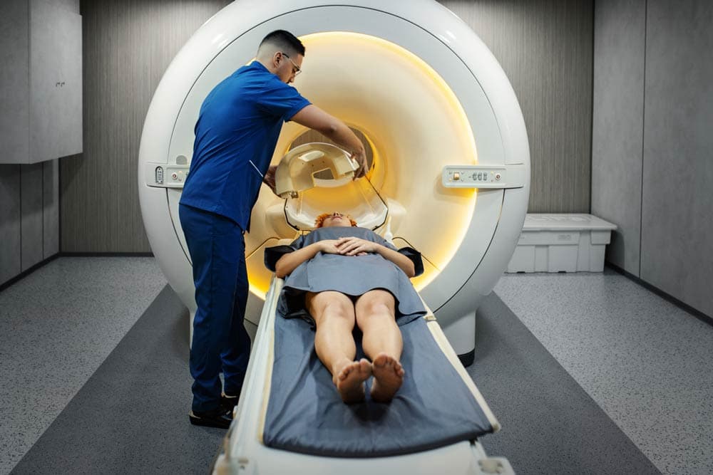
Brain imaging technologies, such as magnetic resonance imaging (MRI), functional MRI (fMRI), and positron emission tomography (PET), have provided valuable insights into the neurobiology of ADHD.
These tools reveal that ADHD is not a character flaw but a neurodevelopmental condition with measurable differences in brain regions and pathways that regulate attention, impulse control, and emotional processing.
Structural Differences in the ADHD Brain
One of the most consistent findings from imaging studies is that certain brain regions are smaller or develop differently in individuals with ADHD.
Shaw et al. (2007) found delayed cortical maturation in children with ADHD, particularly in the prefrontal cortex, the region responsible for executive functions such as planning, organization, and self-control.
Other studies show reduced volume in the basal ganglia and cerebellum, both involved in motor control, reward processing, and timing (Castellanos & Proal, 2012). These structural differences are linked to difficulties with impulse regulation, focus, and coordination.
While these differences tend to lessen with age, they provide strong evidence that ADHD originates in brain development, not environment alone.
Functional Differences: How the Brain Works
Beyond structural changes, brain imaging has shown that functional activity is altered in ADHD.
fMRI studies consistently demonstrate underactivity in the prefrontal cortex and overactivity in the default mode network (DMN). The DMN is a network of brain regions active during rest and mind-wandering but should deactivate during tasks requiring attention.
Sonuga-Barke and Castellanos (2007) described ADHD as involving difficulty suppressing DMN activity, which contributes to distractibility. When individuals with ADHD try to focus, their DMN intrudes, making it harder to sustain attention.
PET scans also reveal differences in dopamine transmission. Volkow et al. (2009) showed that individuals with ADHD have lower dopamine receptor availability in key regions of the reward pathway. This helps explain why everyday tasks may feel less motivating, while stimulating or novel experiences capture attention more easily.
Emotional Regulation and Brain Networks
Brain imaging studies also highlight differences in emotional regulation. The amygdala, which processes emotional responses, shows heightened activity in ADHD, while regulatory regions of the prefrontal cortex show reduced control.
This imbalance contributes to emotional intensity, quick frustration, and sensitivity to rejection (Shaw et al., 2014).
These findings demonstrate that ADHD involves not only cognitive difficulties but also challenges in managing emotions, reinforcing the importance of considering emotional regulation in treatment.
Developmental Trajectories
Longitudinal imaging studies show that brain development in ADHD is not static. Many children eventually "catch up" in cortical thickness, though often at a delayed pace.
Shaw et al. (2007) reported that while cortical maturation lags by several years in children with ADHD, many regions eventually normalize. However, this delay can create significant challenges during crucial academic and social development periods.
This pattern highlights the importance of early intervention, which can help children develop coping strategies while their brains continue to mature.
How Medications Affect the Brain
Imaging studies have also shown how ADHD medications impact brain activity. Stimulant medications increase activation in the prefrontal cortex and normalize dopamine transmission in reward pathways (Rubia et al., 2014).
These changes correlate with improvements in attention, impulse control, and emotional regulation.
This evidence reinforces that medications do not "drug" individuals into submission but instead help restore typical brain functioning, reducing barriers to learning and self-regulation.
Myths Addressed by Brain Imaging
Brain imaging findings help debunk several myths:
- "ADHD isn't real." Imaging shows consistent structural and functional brain differences in ADHD populations.
- "It's just bad behaviour." ADHD brains process information differently, making impulsivity and distractibility symptoms of neurobiology, not defiance.
- "People outgrow ADHD." Imaging reveals persistent differences in brain activity and connectivity, even in adults whose hyperactivity may decline.
By highlighting biological foundations, brain imaging reduces stigma and validates the experiences of those living with ADHD.
Limitations of Brain Imaging
While imaging has provided remarkable insights, it also has limitations. Differences observed in brain scans are often subtle and vary across individuals.
Imaging cannot yet be used as a diagnostic tool for ADHD; diagnosis still relies on clinical assessment and history.
Additionally, many imaging studies use small sample sizes, making replication essential. Researchers caution against oversimplifying findings or using them to predict individual outcomes (Castellanos & Proal, 2012).
The Future of Brain Imaging Research
New techniques, such as diffusion tensor imaging (DTI), which examines white matter connectivity, are expanding our understanding of ADHD.
Large-scale collaborations pooling thousands of brain scans worldwide are helping to clarify patterns and improve reliability.
Future research may allow for more personalized treatment, linking brain imaging markers with responses to specific medications or therapies. The long-term goal is precision medicine, where interventions are tailored to an individual's unique neurobiology.
Final Thoughts
Brain imaging studies provide powerful evidence that ADHD is a neurodevelopmental condition rooted in structural and functional brain differences.
Delayed cortical maturation, altered dopamine signalling, and differences in emotional regulation networks all contribute to the challenges of ADHD.
While imaging cannot diagnose ADHD, it validates lived experiences, reduces stigma, and guides more effective treatments. By understanding what brain imaging reveals, families, educators, and clinicians can approach ADHD with greater compassion and science-based strategies.
References
- 1.Castellanos, F. X., & Proal, E. (2012). Large-scale brain systems in ADHD: Beyond the prefrontal–striatal model. Trends in Cognitive Sciences, 16(1), 17–26. View source ↗
- 2.Rubia, K., Alegria, A., & Brinson, H. (2014). Brain abnormalities in attention-deficit hyperactivity disorder: A review. Revista de Neurología, 58(S01), S3–S16. View source ↗
- 3.Shaw, P., Eckstrand, K., Sharp, W., Blumenthal, J., Lerch, J. P., Greenstein, D., … & Rapoport, J. (2007). Attention-deficit/hyperactivity disorder is characterized by a delay in cortical maturation. Proceedings of the National Academy of Sciences, 104(49), 19649–19654. View source ↗
- 4.Shaw, P., Stringaris, A., Nigg, J., & Leibenluft, E. (2014). Emotion dysregulation in attention deficit hyperactivity disorder. American Journal of Psychiatry, 171(3), 276–293. View source ↗
- 5.Sonuga-Barke, E. J., & Castellanos, F. X. (2007). Spontaneous attentional fluctuations in impaired states and pathological conditions: A neurobiological hypothesis. Neuroscience & Biobehavioral Reviews, 31(7), 977–986. View source ↗
- 6.Volkow, N. D., Wang, G. J., Kollins, S. H., Wigal, T. L., Newcorn, J. H., Telang, F., … & Swanson, J. M. (2009). Evaluating dopamine reward pathway in ADHD: Clinical implications of imaging studies. American Journal of Psychiatry, 166(5), 564–573. View source ↗